"A severe lateral ankle sprain is not just a temporary swelling of the foot; it represents a violent mechanical failure of the primary stabilization ligaments. Restoring an elite soccer player to competitive pivots demands precise ligament remodeling, progressive tissue loading, and rigorous proprioceptive retraining."
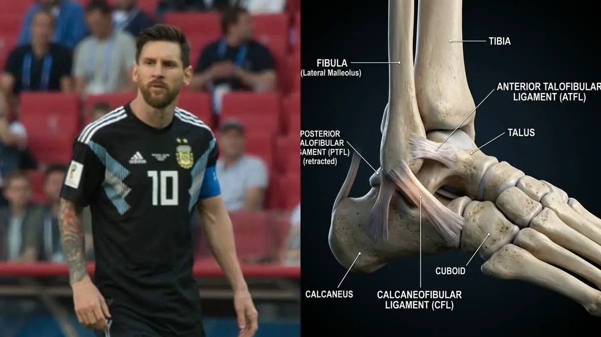
In the final match of the Copa America tournament, millions of football fans watched in shock as Lionel Messi was forced off the pitch in tears. Replays and subsequent close-ups revealed a massive, balloon-like swelling on the lateral side of his right ankle. The diagnosis was confirmed as a severe lateral ankle ligament sprain, which sidelined the Argentine legend for several weeks. This high-profile injury highlighted the vulnerability of the human ankle during the high-speed pivots and cutting maneuvers characteristic of elite-level soccer.
While an ankle sprain is one of the most common sports injuries, the rehabilitation process for an elite athlete is complex. It involves much more than just resting and applying ice. In this clinical biomechanical analysis, we will explore the precise anatomy of lateral inversion sprains, dissect the grading criteria of ligament tears, and detail the multi-stage rehabilitation protocol designed to safely return athletes to high-impact cutting sports.
The Anatomy and Biomechanics of Lateral Inversion Sprains
The ankle joint is a highly complex structure, consisting of the talocrural joint (the primary hinge joint between the tibia, fibula, and talus) and the subtalar joint. Lateral stability is maintained by three distinct ligaments, which are stretched or torn during an inversion sprain:
- Anterior Talofibular Ligament (ATFL): This is the weakest of the three ligaments and the most commonly injured. It runs horizontally from the front of the fibula to the talus, preventing anterior sliding of the foot.
- Calcaneofibular Ligament (CFL): This ligament runs vertically from the lateral malleolus of the fibula to the calcaneus (heel bone), providing primary stability against inversion when the ankle is in a neutral or dorsiflexed position.
- Posterior Talofibular Ligament (PTFL): The strongest of the lateral ligaments, running from the posterior aspect of the fibula to the talus. It is rarely injured, except in cases of complete ankle dislocation.
An inversion ankle sprain occurs when the foot rolls inward while the ankle is in a state of plantarflexion (pointed downward). This combination of plantarflexion and inversion stretches the lateral ligaments beyond their physiological limits, beginning with the ATFL. If the force continues, the CFL is also damaged, potentially leading to joint instability and secondary tissue trauma.
- Lateral ankle sprains account for approximately 70% to 80% of all ankle injuries in field sports.
- Recurrence rates among elite soccer players can be as high as 30% to 40% if proprioceptive training is omitted.
- A Grade II ligament tear reduces local mechanical stability, requiring up to 6 to 8 weeks for the ligament fibers to remodel and regain tensile strength.
- Elite players perform an average of 700 to 800 cutting maneuvers per match, placing repetitive, high-velocity lateral shear forces on the healing ligaments.
Clinical Assessment and Determining Injury Severity
Clinically, ankle sprains are classified into three distinct grades based on the extent of tissue damage and the resulting joint laxity:
Grade I (Mild): Microscopic tearing of the ligament fibers (usually isolated to the ATFL) with mild swelling and tenderness, but no structural joint instability. The patient can bear weight with minimal pain.
Grade II (Moderate): Partial tearing of the ATFL and CFL. This results in moderate-to-severe swelling, localized bruising (ecchymosis), significant pain, and mild joint laxity during stress tests. Weight-bearing is painful and difficult.
Grade III (Severe): Complete rupture of the ATFL and CFL, and occasionally the PTFL. This leads to massive swelling, widespread bruising, severe pain, and significant mechanical joint instability. The patient is unable to bear weight without extreme discomfort.
"In Messi's case, the visible swelling was immediate and severe, indicating a Grade II or Grade III injury with significant bleeding from the microvascular structures surrounding the ligaments. In elite rehabilitation, our first priority is to rule out associated injuries, such as fractures of the lateral malleolus or fifth metatarsal, and syndesmotic (high ankle) sprains. We use the Ottawa Ankle Rules to determine the need for X-rays, followed by high-resolution MRI to map the exact integrity of the ATFL and CFL fibers."
The Multi-Stage Elite Soccer Rehabilitation Protocol
Reclaiming lateral stability and explosive power requires a structured, progressive loading protocol that respects the phases of biological tissue healing.
- 1Phase 1: Protection and Inflammation Control (Days 1–7)Apply the POLICE principle (Protection, Optimal Loading, Ice, Compression, Elevation). Protect the ankle using a semi-rigid brace that allows sagittal movement (plantarflexion and dorsiflexion) but restricts inversion. Begin early, pain-free optimal loading to stimulate ligament alignment. Use intermittent compression therapy and active calf pumping to clear fluid from the joint cavity.
- 2Phase 2: Restoring Mobility and Strength (Weeks 2–4)As pain and swelling subside, focus on restoring full dorsiflexion range of motion, which is crucial for running mechanics. Perform joint mobilizations, calf stretches, and active ankle circles. Begin isometric eversion and inversion exercises, progressing to eccentric strengthening using resistance bands. Start double-leg balance exercises on stable surfaces, progressing to single-leg balance.
- 3Phase 3: Neuromuscular and Proprioceptive Retraining (Weeks 5–6)Re-educate the ankle's mechanical receptors to prevent chronic instability. Perform balance work on unstable surfaces (wobble boards, foam pads) while throwing and catching a ball. Introduce dynamic agility drills, linear jogging, and light jumping (plyometrics). Incorporate closed-chain strengthening exercises like single-leg squats and lunges.
- 4Phase 4: Sport-Specific Conditioning and Pivoting (Weeks 7+)Introduce high-velocity soccer-specific movements, including lateral cutting, acceleration/deceleration drills, and ball control exercises. The athlete must demonstrate equal single-leg hop distances and vertical jump heights compared to the uninjured side. Return to full training with a protective tape job or low-profile brace for the first few competitive matches.
The Patient: Marcus, a 22-year-old collegiate soccer player who sustained a Grade II lateral ankle sprain during a match, exhibiting severe lateral joint swelling.
The Mistake: Marcus rested the ankle for three weeks, waiting for the pain to disappear. When he returned to play without rehabilitation, he suffered a recurrent sprain within ten minutes, accompanied by chronic lateral ankle pain.
The Solution: We initiated a structured proprioceptive program, focusing on eccentric evertor strengthening, dynamic balance drills, and lateral cutting mechanics under video analysis.
The Outcome: After six weeks of active physical therapy, Marcus returned to competitive play with full ankle stability, no pain, and has remained injury-free for two full seasons.
Synergy in Lower Extremity Rehabilitation
Ankle injuries often alter the biomechanics of the entire leg, leading to secondary issues in the calf, knee, and foot. For a detailed guide on restoring mechanical stability and ankle movement, see our foundational guide on ankle sprains and proprioception. If you are also managing chronic tendon pain, study our protocols for Achilles tendinitis rehabilitation. Additionally, for a comparative look at high-energy ankle fixation and return-to-sport timelines, read our motorsport case study on Alexander Rossi's Indy 500 crash and ankle recovery.
Lionel Messi's recovery from his Copa America ankle injury demonstrates the effectiveness of modern sports medicine and physical therapy. By progressing systematically through swelling management, strength building, and intensive proprioceptive retraining, athletes can recover from severe ligament sprains and return to their sports with confidence. The key is to respect the biological healing timeline while actively restoring the neuromuscular pathways that protect the joint during explosive movements.
Featured image: Clinical side-by-side composite showing Lionel Messi (left) and an anatomical 3D rendering of the human ankle joint showing lateral ligaments (right). Created for AyurPhysio editorial use. Wikimedia Commons attribution: Lionel Messi image by Екатерина Лаут / Ekaterina Laut licensed under CC BY-SA 3.0. Modified by cropping and compositing.
Dr. Dhanushika Dilshani
Expert Ayurvedic Wellness Doctor. Specialized in modern holistic wellness, optimizing dermal resilience, cosmetic radiance, and systematic diagnosis driven by traditional and evidence-based medical logic.
Medical Disclaimer
The information provided by AyurPhysio is for general educational and informational purposes only. It is not intended as a substitute for professional medical advice, diagnosis, or treatment. Always seek the advice of your physician or other qualified health providers with any questions you may have regarding a medical condition. Never disregard professional medical advice or delay in seeking it because of something you have read on this website.
Related Healing Guides
View All Guides →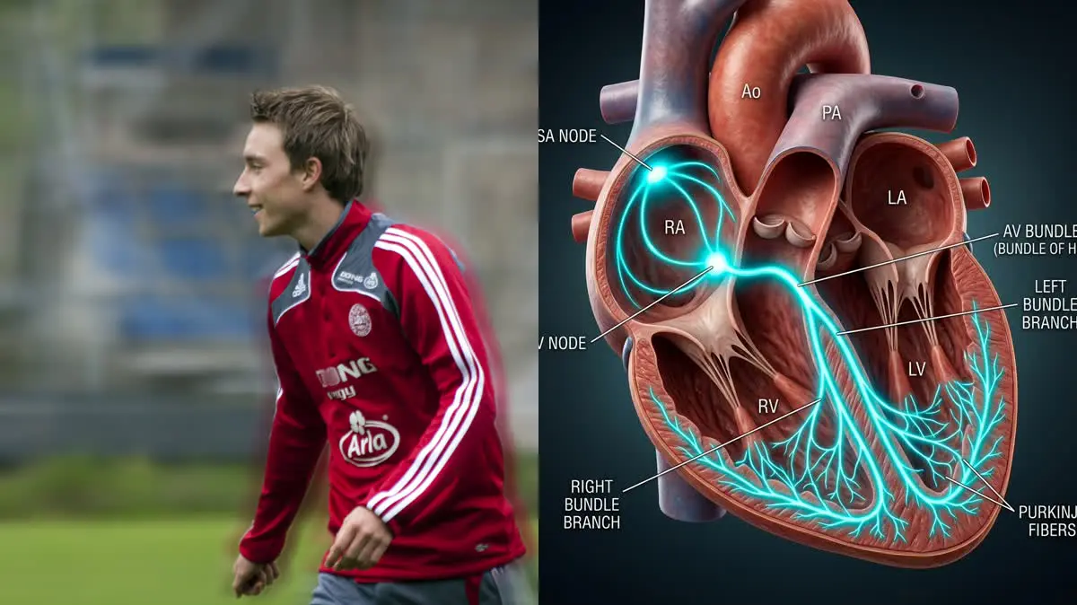
Christian Eriksen's Cardiac Arrest and ICD Return to Play: The Biomechanics of Cardiorespiratory Reconditioning and Athletic Heart Rehabilitation

Selena Gomez's Lupus and Kidney Transplant: An Integrative Ayurvedic Approach to Systemic Autoimmunity, Dushi Visha, and Ojas Restoration